 The Clarinet BBoard The Clarinet BBoard
|
Author: SunnyDaze
Date: 2019-07-15 01:47
Hi,
I just had a thought today and I wondered if I could ask about it?
I'm an adult learner on the clarinet, but professionally I am mainly a microscopist, and I have my own microscope setup at home (I'm mostly a stay-at-home Mum just now). My microscope is a brightfield setup, which means that it just takes photos like a normal camera but at very high magnification.
I've been watching videos on youtube about how people adjust their reeds and I wondered if there is any part of the reed that it would be interesting for you all to see under huge magnification? If there isn't, don't worry about it, but I just thought I would offer, in case it would be useful. I'm a botanist, so wooden reeds are well within the range of what I am meant to be able to do.
The best photos that I can take will magnify a subject that is about 2.5mm by 3.5mm so that it can be printed at high resolution at A3 paper size. I can also theoretically photograph a subject that is 0.5mm by 0.7mm and print it to A3 but I haven't actually tried that yet. I can also do bigger subjects.
I wondered, for example, would it be useful to look at the sharp end of a clarinet reed and see how one brand differs from another? Or would it be useful to see how the wood changes when it is smoothed down, as people recommend doing in some videos that I have seen. I could have a go at pretty much anything, if you think it would be useful.
Thanks for thinking about it!
Sunny
|
|
Reply To Message
|
|
Author: johng ★2017
Date: 2019-07-15 02:02
I think any pictures you could get would be great to see. As you suggest, the thin tip would be the most interesting. Great suggestion, thanks!
John Gibson, Founder of JB Linear Music, www.music4woodwinds.com
|
|
Reply To Message
|
|
Author: SunnyDaze
Date: 2019-07-15 02:18
Hi John,
Thanks, I will have a go then and see what I get. :-)
Sunny
|
|
Reply To Message
|
|
Author: kdk
Date: 2019-07-15 05:31
It would be interesting to see the differences among strengths of a given reed brand and model. The difference in strength, for example, between a #3 and a #4 Vandoren V.12 is supposed to be the result of the greater density of the cane the stronger (higher number) reed is made from. It would be interesting to see what that actually looks like.
It would also be very instructive to see how a reed's structure changes over one, two or three wetting/drying cycles, as from the first time you play on a reed, through the next two or three playings. We all know a reed's response changes over these first few wet/dry cycles - it's the reason for all the various routines players develop to "break in" new reeds. But most of us just intuit what physical changes actually take place.
Would your equipment be able to make microbes visible growing in the cane? They might be too small or need some additional technique (dye?) to make them visible.
I've seen fairly detailed photos of reeds in articles before, but I don't think at the level of magnification and resolution you're suggesting.
|
|
Reply To Message
|
|
Author: clarnibass
Date: 2019-07-15 08:48
A bit off topic maybe but... I also do some extreme macro photography. What magnification are your lenses and what camera do you use?
My current setup is up to x10 and using an APSC 24MP camera, this means that a 2.40mm x 1.60mm rectangle would fill the frame. This could print, at native resolution (without software or printer enlarging or reducing size), high quality of 300 DPI, at about 51cm x 34cm (roughly 20" x 13").
Of course many things can affect the result. The printer, type of paper, aperture used (usually fixed for telescope lenses), etc.
I also found that 240 DPI or even 180 DPI can look excellent. Depending on distance, can be indistinguishable.
I've printed to 1.50m x 1.00m at 240 DPI and it was good.
Also projected on a screen at just FHD resolution even much lager and it was good.
There is a whole world to learn about printing...
Do you use a single frame or do you stack? I'm guessing you know the extremely thin depth of field you get at these magnifications. I'm wondering how it works with your camera and microscope?
|
|
Reply To Message
|
|
Author: ruben
Date: 2019-07-15 09:11
Fascinating proposal, Sunny! Just to be pedantic, cane is not, strictly speaking, wood. It would be interesting to know if there is a structural difference between cane coming from different areas: cane from the Var in the South of France as opposed to that coming from Argentina or Australia for example. There must be a difference in the fibers. My theory is that the constant wind in the Var (le Mistral) makes the cane more fibrous. Good luck with your research!
rubengreenbergparisfrance@gmail.com
|
|
Reply To Message
|
|
Author: Bob Bernardo
Date: 2019-07-15 10:07
I actually have a few microscopes but unable to take photos.
I have done a lot of studies on cane from when I worked at Rico for for many years doing R&D (Research and Development) so this is very cool. After Rico I also did some work; studies at CalTech.
Please email me when you have some time and lets see what we can do together to advance this subject. There is also a need for less power, perhaps 10X or so. I'm not sure if you are talking about 100x to 400x or even higher. However, I've done this and it would be cool to put our heads together on this project; if interested. Send me your phone number if interested. There are a lot of secrets here to be seen under power and also takes away some of the guess work and mysteries of what makes good and bad reeds from the very start of plantations soil and much more to the final cut of the good or bad reed. This includes things like why do reeds die after 2 minutes or last for 2 months. These are all of the answers I have complete knowledge of. I could be one of the very few people in the world that has the information and it would be a blast to put this into photography.
savagesax@aol.com
Designer of - Vintage 1940 Cicero Mouthpieces and the La Vecchia mouthpieces
Yamaha Artist 2015
|
|
Reply To Message
|
|
Author: SunnyDaze
Date: 2019-07-15 12:52
Hi,
Thanks so much for all of your encouraging responses. It's great to think that my microscope might be useful for the clarinet community. It's really exciting to know that there are other specialists and microscopists out there too. I reckon we could have some fun if we work together.
Bob - it sounds as though you have done a lot of this before, but perhaps the results were industry secrets. Is that right? Maybe I could do the public version in a small way, if your work is very secret, very advanced, and not to be shared online?
Clarnibase, your setup sounds great, and very much like mine. That's brilliant that there are two of us the same here. It's always better with a group working together. :-)
For the technical detail, a description of my setup for a general audience is given in the video below (Please do laugh at my accent btw. The funding body asked me to make the videos as a condition of my funding for the microscope objectives. Even I still laugh at the videos.) :
https://www.youtube.com/watch?v=5IGpcFpEbxM
The full detail is here of the setup is here:
http://chlorophyllosophyimages.blogspot.com/2017/12/focus-stacking-setup.html
The equipment that I use is a Canon 5d MkII camera, a Olympus Zuiko 200mm prime lens and a series of Mitutoyo microscope objectives (x5, x10, x20, x50). I have a Canon MP-E lens for bigger specimens and an Olympus Zuiko 2:1 lens for even bigger ones.
My area of special interest is gametophyte ferns, but I reckon I could have a go at clarinet reeds too and see how it goes. The funding that paid for the microscope objectives was for plant science research, and I'm pretty certain that clarinet reeds count as applied plant science research.
I can't start on it quite immediately because my kitchen wall is being demolished this week, and my microscope is just outside the kitchen door, but hopefully next week (assuming that the house is still standing.) :-)
I really like the idea of first photographing the thin tip of the different strengths of one brand of reed. I will aim for that first. I have several strengths of classic vandoren reed, so I am well set up for that.
The idea of photographing new reeds over several initial wet-dry cycles sounds great too. Now that I am learning grade 3 I am realising that reeds are really really important for getting the right sound and managing the tricky technical bits, so I will be very interested to learn about the importance of reed handling and breaking in. I will put that down for experiment number 2.
I wonder if experiment number 3 might be to look at the difference between the tip of the one reed from the box that works perfectly and the other that doesn't work at all? It seems to me that one is always just too hard, but I may be quite wrong.
Experiment number 4 in my head is to figure out what I need to do to make the non-working reed work, and whether the photos can help with that.
Yay!
Sunny
|
|
Reply To Message
|
|
Author: clarnibass
Date: 2019-07-15 13:58
Yes, our setups are pretty similar. I use both FF and APSC cameras, but usually the APSC which allows lower magnification to fill the frame. I also use Mitutoyo objectives, but only up to x10 (equivalent to x15 on FF, not considering resolution). I use an electronic rail too, a pretty similar flash setup and Zerene for stacking.
I've done some macro with clarinets parts, like rusty springs, etc. but not with the highest magnification, only about x2 or less so far. I haven't tried a reed.
For lower magnification than x5 I use mostly coupled reversed lens, mine happen to be Nikon, or just plain macro lenses for x1 or less.
Maybe I'll try the reed too.
|
|
Reply To Message
|
|
Author: SunnyDaze
Date: 2019-07-15 14:06
Hi Clarnibass,
That's brilliant that you have the same setup. I'd be really interested to see what you get if you try reed photography. :-)
What is the minimum effective step size on your rail? I was aiming for 1 micron when I built my rail, but in practice I've never needed to go below about 10.
Sunny
|
|
Reply To Message
|
|
Author: Ken Lagace
Date: 2019-07-15 17:33
I would like to see the difference in reed cane from different parts of the world. If interested, I could search my collection and send you some.
|
|
Reply To Message
|
|
Author: SunnyDaze
Date: 2019-07-15 17:43
Hi Ken,
That's a very interesting thought. I suppose the cane may be quite different from different places.
I will have a little go at taking some photos once I get my house together next week, and if I can make a decent job of it then that would be lovely to have have a go at photographing some of your reeds.
Thanks :-)
Sunny
|
|
Reply To Message
|
|
Author: Tony Pay ★2017
Date: 2019-07-15 19:01
There is a reed adjustment technique promoted by Tom Ridenour that he calls 'the ATG system', the letters standing for 'Against The Grain'.
The idea is that sanding the top surface of a reed from tip to butt, rather than the other way, deals with the fibres of the cane more effectively. It's like shaving 'against the grain', when removing hairs from your skin. The razor cuts the hairs off at the bottom rather than tending to flatten them against the skin.
I've always thought the idea intuitively sound, and use the technique myself; but it would be nice to have some blown-up pictures, sideways on, showing the difference in the reed's behaviour under 'butt to tip' and 'tip to butt' sanding.
A research project?
Tony
|
|
Reply To Message
|
|
Author: SunnyDaze
Date: 2019-07-15 19:31
Hi Tony,
Thanks, yes that sounds good and is probably the easiest of the suggested experiments. I'll put it on the list. :-)
Thanks!
Sunny
|
|
Reply To Message
|
|
Author: ruben
Date: 2019-07-15 19:37
Ken: Do you have any cane from Cuba? Ages ago, Russel Procope of the Duke Ellington orchestra told me that the world's best cane was Cuban and he lamented
the fact that because of the Cuban embargo, it was no longer available.
rubengreenbergparisfrance@gmail.com
|
|
Reply To Message
|
|
Author: SunnyDaze
Date: 2019-07-15 21:27
There seem to plant science publications about this. Yay!
https://www.jstor.org/stable/42764997?Search=yes&resultItemClick=true&searchText=Arundo&searchText=donax&searchUri=%2Faction%2FdoBasicSearch%3FQuery%3DArundo%2Bdonax%2B%26amp%3Bacc%3Doff%26amp%3Bwc%3Don%26amp%3Bfc%3Doff%26amp%3Bgroup%3Dnone&ab_segments=0%2Fdefault-2%2Fcontrol&refreqid=search%3Afc44d995d2e84a6e545faedb473f0e19&seq=1#page_scan_tab_contents
https://www.jstor.org/stable/4287997?Search=yes&resultItemClick=true&searchText=Arundo&searchText=donax&searchUri=%2Faction%2FdoBasicSearch%3FQuery%3DArundo%2Bdonax%2B%26amp%3Bacc%3Doff%26amp%3Bwc%3Don%26amp%3Bfc%3Doff%26amp%3Bgroup%3Dnone&ab_segments=0%2Fdefault-2%2Fcontrol&refreqid=search%3A25b2f9c12f656a18bc11d6be50b5a007&seq=1#page_scan_tab_contents
|
|
Reply To Message
|
|
Author: SunnyDaze
Date: 2019-07-15 21:29
Maybe Bob Bernardo knows the whole story and could tell us which photos are really worth taking?
|
|
Reply To Message
|
|
Author: jeeves
Date: 2019-07-17 05:37
Following up on http://test.woodwind.org/clarinet/BBoard/read.html?f=1&i=475597&t=475597, discussing the degradation of Legere reeds, if you have a new Legere reed and a worn-out Legere reed, I think it would be interesting to compare the two and see if there's any evidence of cracking/crazing in the worn-out reed.
|
|
Reply To Message
|
|
Author: SunnyDaze
Date: 2019-07-17 11:12
Hi David,
That book sounds great. Is there any chance that you could find the name of it and maybe I could get it on interlibrary loan?
Thanks!
Jen
|
|
Reply To Message
|
|
Author: SunnyDaze
Date: 2019-07-17 11:13
Hi Jeeves,
I don't have any Legere reeds, but I could put that on the list.
Thanks!
|
|
Reply To Message
|
|
Author: Bob Bernardo
Date: 2019-07-17 21:31
Yes there are a lot of secrets. This is all that I wish to say right now and all that I wish to moment on.
Designer of - Vintage 1940 Cicero Mouthpieces and the La Vecchia mouthpieces
Yamaha Artist 2015
|
|
Reply To Message
|
|
Author: clarnibass
Date: 2019-07-17 21:56
Sunny, I think my rail can theoretically do 1 micron steps. The controller has that option but I've never tried that and don't know if it can do that accurately. I guess I could test this with a puppitast but I don't have high enough magnification to test that with a stack. I've never needed less than a 0.015mm step and even that is rare. The rail itself uses linear rails and originally made for machines or maybe robotics, etc. which is why it's precise and (relatively) affordable.
|
|
Reply To Message
|
|
Author: SunnyDaze
Date: 2019-07-18 09:09
Hi Bob,
Thanks, yes, I completely understand. Maybe we can do a bit of public research here and the private stuff can stay private, as it should. :-)
Thanks!
|
|
Reply To Message
|
|
Author: Bob Bernardo
Date: 2019-07-19 07:23
Hi SunnyDazy
When or if you are ready you have my email addy. I'd be interested in a publication type study. Copyright situation which could be very helpful to the clarinet and sax world.
There are so many myths and really dumb comments about how people treat their reeds and adjust their reeds. I often shake my head in disbelief. When I try to help people; often giving them some free reeds in the past, they don't get it.
An example is keeping wet reeds in a zip lock plastic baggy then mold develops. This can cause all sorts of harmful body issues. Yet I see repeat threads about this. Well needless to say I DEEPLY care about the health of fellow players. I could go on and on. People never wash their mouthpieces and clean their horn for years and years. The clarinet swaps look like something from the 1940's.
Designer of - Vintage 1940 Cicero Mouthpieces and the La Vecchia mouthpieces
Yamaha Artist 2015
|
|
Reply To Message
|
|
Author: SunnyDaze
Date: 2019-07-19 11:36
I just noticed that my local shop have very hard reeds on special clearance sale just now so I bought no. 5 strength versions of the three Vandoren kinds that I already have in 2.5. That should get me off to a good start. :-)
|
|
Reply To Message
|
|
Author: SunnyDaze
Date: 2019-07-20 11:25
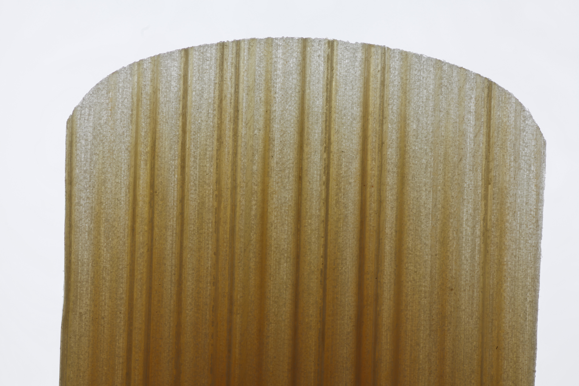
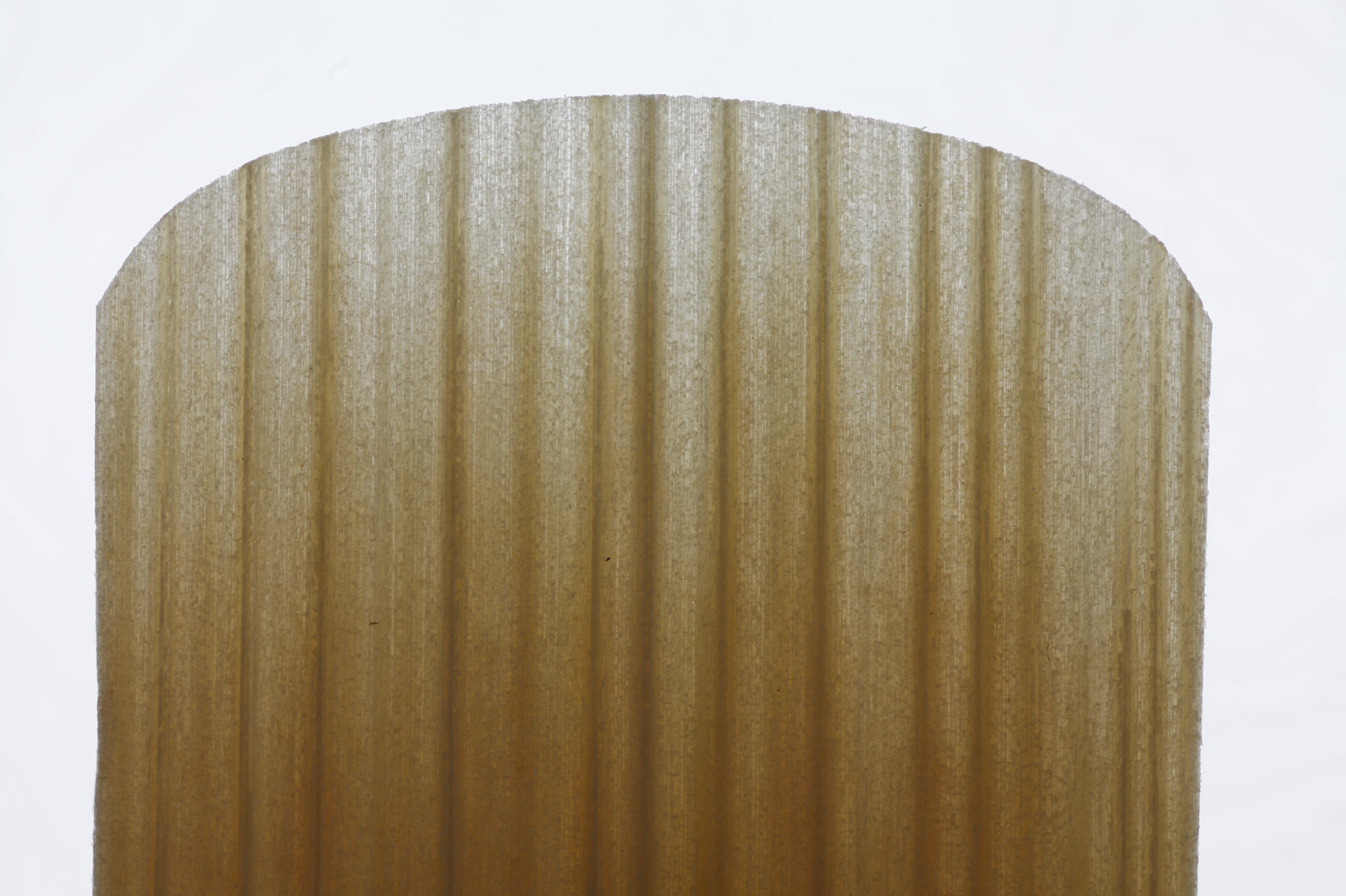
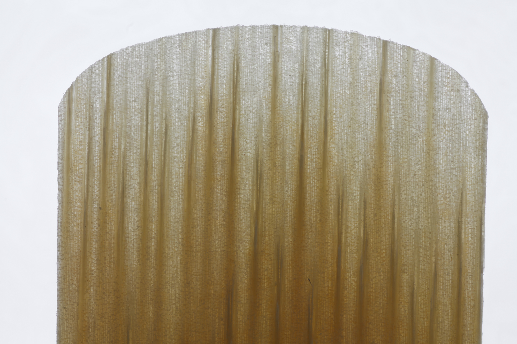
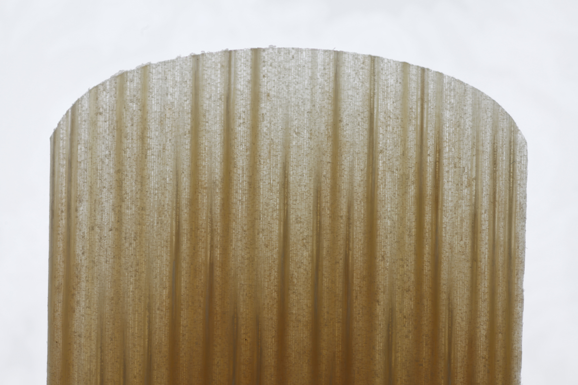
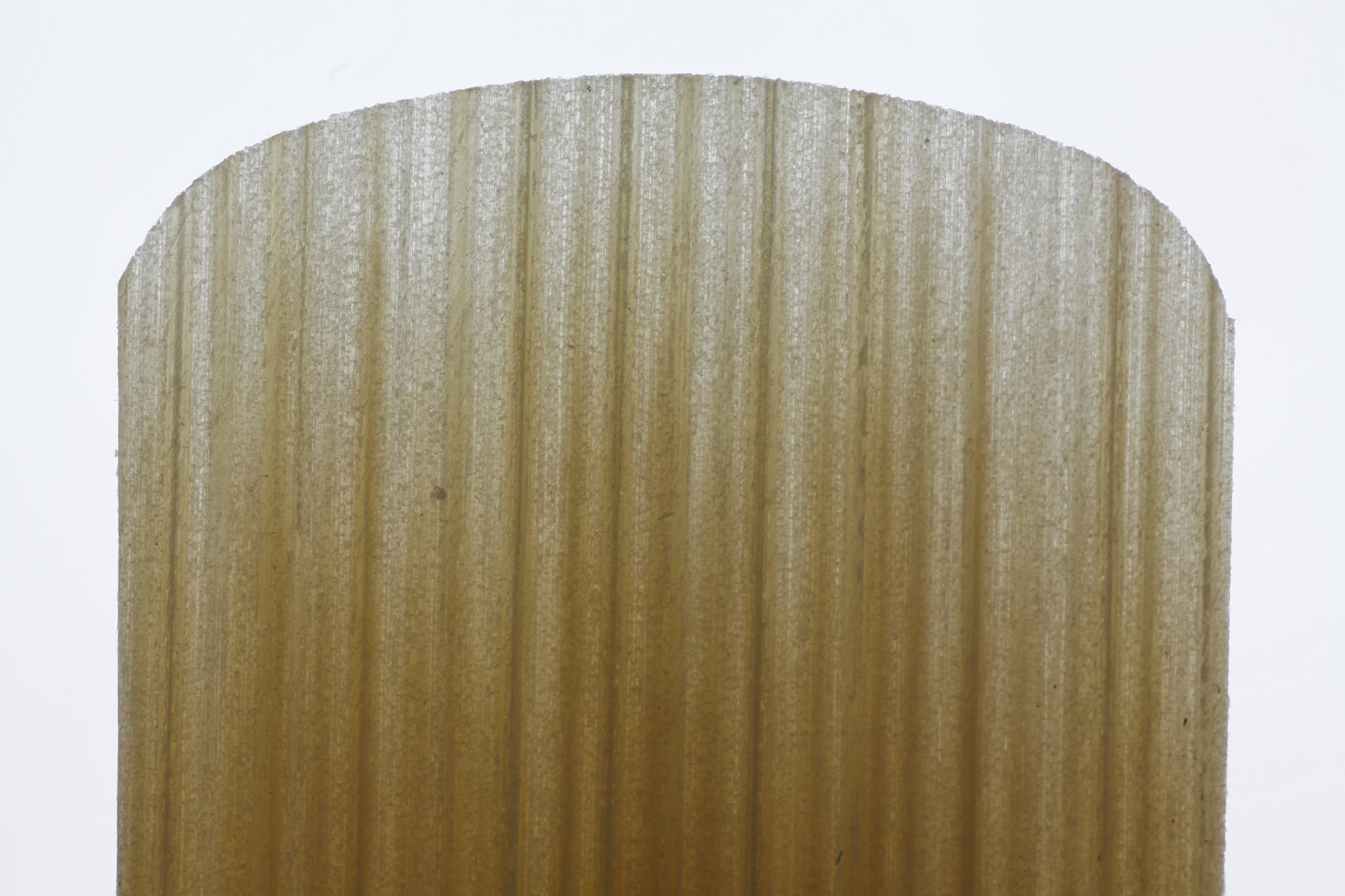
Attachment: 1.5-strength-67-slices.jpg (177k)
Attachment: 3-strength-67-slices.jpg (171k)
Hi,
Here are my first couple of photos. They are not super-exciting. :-)
They are of the sharp end of a well used 1.5 reed and a once-used 3 reed. I'll not say which brand, because I've written to the company to ask if they happy to have close-ups posted and haven't heard back yet.
Sunny
|
|
Reply To Message
|
|
Author: Ken Lagace
Date: 2019-07-20 19:54
These are really cool. What are the tiny threads that seem to be in all images?
|
|
Reply To Message
|
|
Author: SunnyDaze
Date: 2019-07-20 20:59
Attachment: composite-1small.jpg (1764k)
Hi Ken,
I'm not sure, but the humorist in me wonders if that is the bit that matters. :-)
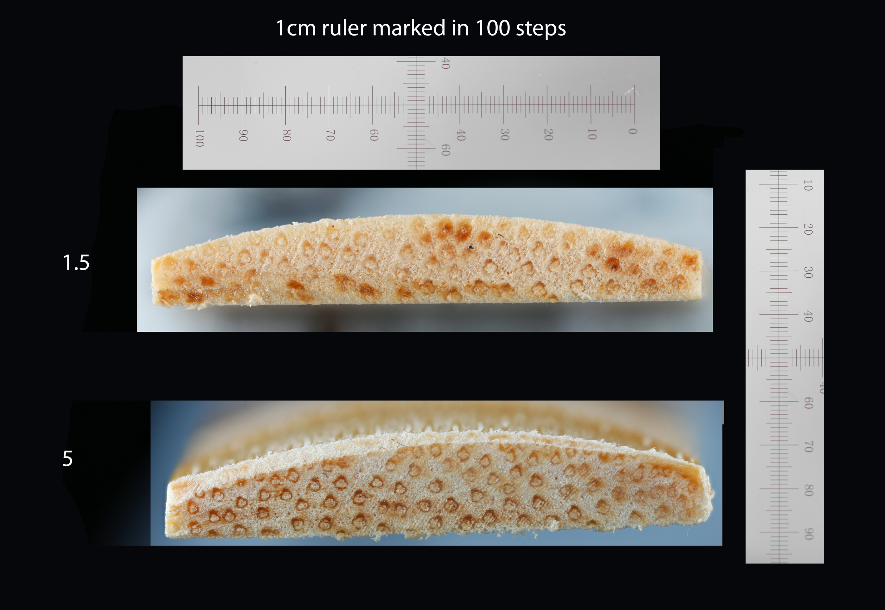
One of the staff at my local shop suggested the attached photo, which is the 5 and 1.5 reeds cut across in the heart of the reed. I cut at 26mm from the tip with a demel multidrill with an angle grinder bit attached.
The thicker reed has a little ledge at the top and the top of the reed is at the top of that ledge rather than the bottom.
They are really clearly thicker at that point. It's good to know that there is something to see. I was starting to wonder if it was all magic.
Sunny
|
|
Reply To Message
|
|
Author: SunnyDaze
Date: 2019-07-20 21:34
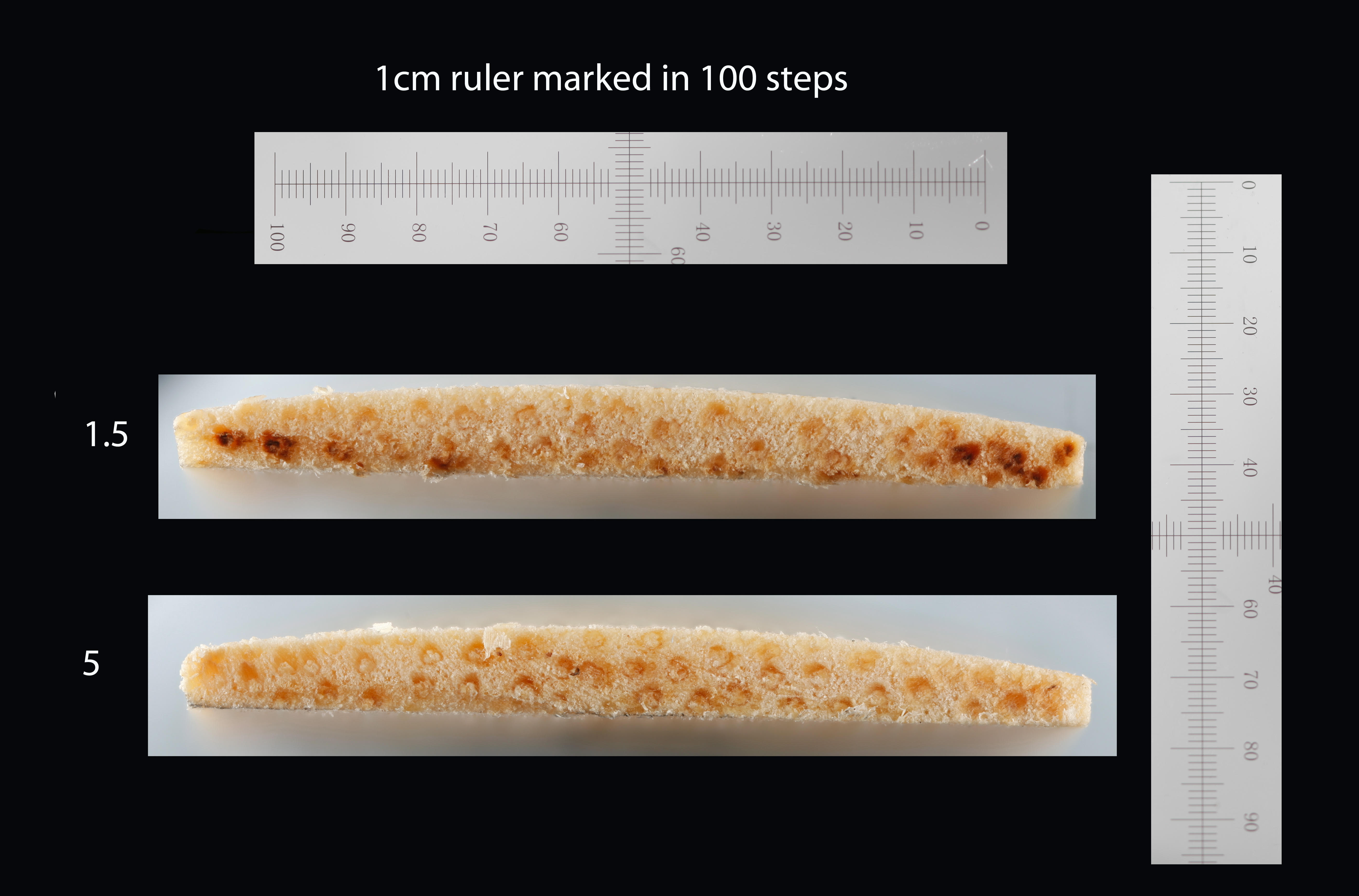
Attachment: compositesmall.jpg (1657k)
Hi,
Here is another composite. The reed was cut through in the same way, but 18mm from the tip. The difference is much less apparent here.
That's me done for today. :-) Thanks for looking.
Sunny
|
|
Reply To Message
|
|
Author: kdk
Date: 2019-07-22 05:31
Are the 5 and the 1.5 the same brand and the same model? That the #5 is actually thicker would put the lie to what at least some reed manufacturers claim, that the reeds are all cut from the same blanks (same thickness) and are sorted into strengths after they are cut. The difference is supposed to be the hardness or amount of flexibility of the cane itself. But you'd need to be comparing a V12 #1.5 to a V12 #5 or the equivalent in other brands.
Even so, there is, I've found, a fair amount or variability in the thickness even of reeds of the same strength and model. Measuring at the end of the bark where the vamp begins, V.12s ("thick blanks") can be anywhere from 3.05 mm or 3.1 mm all the way up to 3.6 mm or even 3.7 mm. Vandoren Traditionals ("Blue box/Thin blanks") are always less than 3 mm at the same point, some as thin as 2.5 mm. So it would be very likely that any two reeds, even if you were comparing two V.12s of different strengths, would not be the same thickness.
Karl
|
|
Reply To Message
|
|
Author: SunnyDaze
Date: 2019-07-22 14:06
Hi Karl,
I just heard back from Vandoren that they are happy for me to make the information public (Very good of them to say that), so I can now say that they are both Classic Vandoren reeds (blue box).
I only did one of each, but I agree, they do seem to be very different thicknesses. The composition of the plant material does seem very different as well though. In the thick reed the vascular bundles are much more distinct, as though they are more mature, and might have much thicker, stronger cell walls, which would then stiffen the reed.
Vascular bundles are the pipes that run up and down the plant to transport water upwards from the roots and sugar downwards from the leaves. There is a photograph of a cross section of Arundo donax stem in figure A here:
https://www.researchgate.net/figure/Cross-sections-of-Arundo-donax-L-stem-and-leaf-A-stem-B-magnified-part-of-stem-C_fig1_322283685
Arundo donax is the grass from which a clarinet reed is made. This cross section looks very much, to me, like the photo of the stronger reed.
Sunny
|
|
Reply To Message
|
|
Author: SunnyDaze
Date: 2019-07-22 14:32
My hope in doing this is to learn what I need to adjust in my reeds to make them perform well in particular styles of music, but I think I'm quite a long way from figuring that out. :-)
|
|
Reply To Message
|
|
Author: Tony Pay ★2017
Date: 2019-07-22 17:32
SunnyDaze wrote:
>> My hope in doing this is to learn what I need to adjust in my reeds to make them perform well in particular styles of music, but I think I'm quite a long way from figuring that out. :-) >>
...as is the rest of the world.
The thing is, guessing how an object will vibrate is a difficult problem, not to be reduced to characterising its physical dimensions, including variables like fibre density.
I used to be surprised that the bows of string instruments weren't easily made out of synthetic materials, even though I could see why that wasn't possible for the instruments themselves. When I found out that the wood of a bow vibrates as you use it – even how you hold a bow makes a difference – I was less surprised.
Tony
|
|
Reply To Message
|
|
Author: SunnyDaze
Date: 2019-07-22 17:52
Yes I see what you mean. That would explain a lot.
I have got far enough now to realise that I do better with one reed for a tune that is all articulated high notes and another that is all crossing the break legato. It seems as though narrowing it down and learning more about reeds would be a good idea.
Do you think it's a good idea to write to the people who made the mouthpiece and ask what they recommend? I play with a J&D Hite D mouthpiece.
Thanks!
|
|
Reply To Message
|
|
Author: SunnyDaze
Date: 2019-07-22 18:02
I have tried writing to the manufacturers. I will wait and see what happens.
|
|
Reply To Message
|
|
Author: johng ★2017
Date: 2019-07-22 20:13
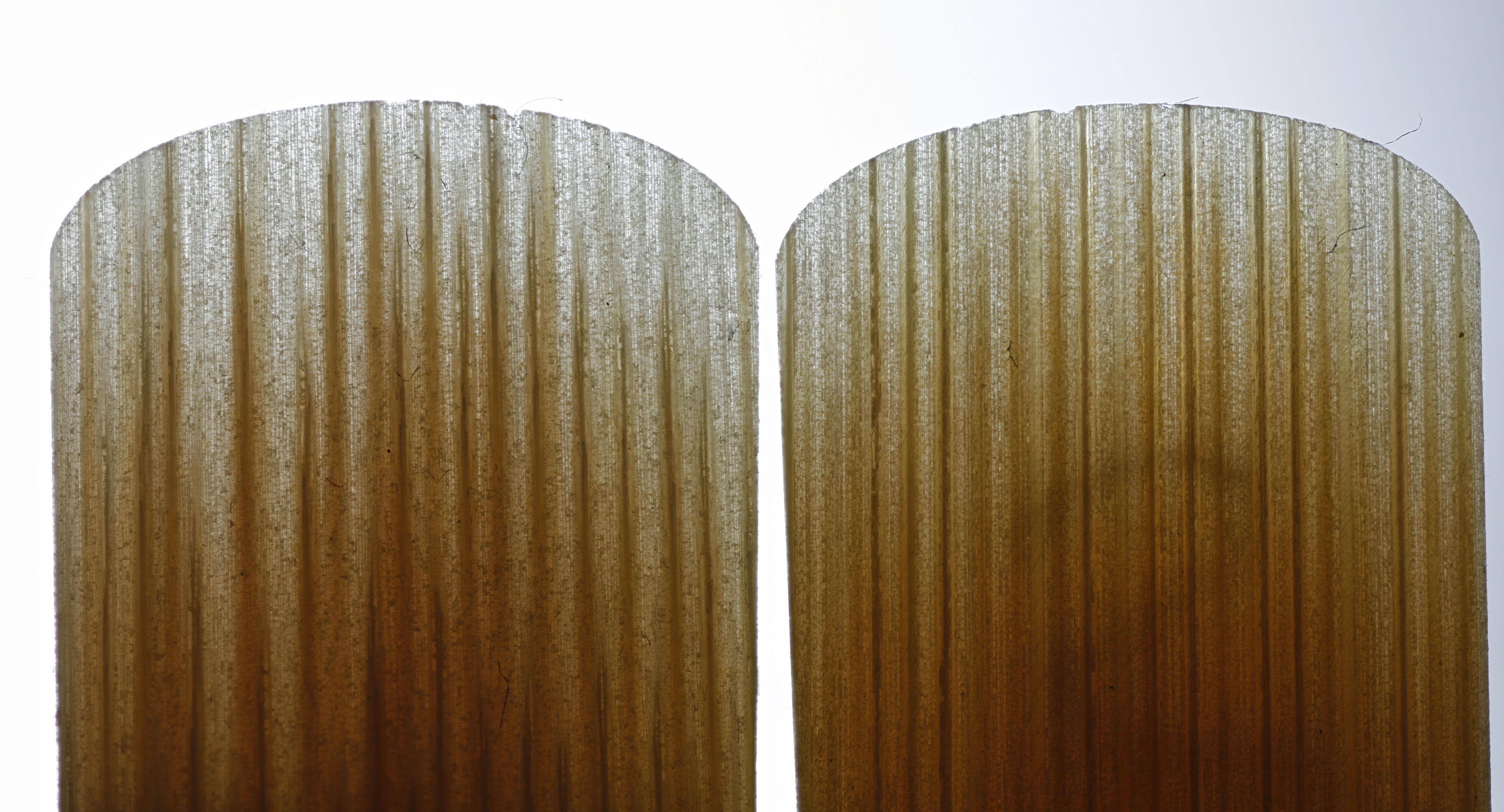
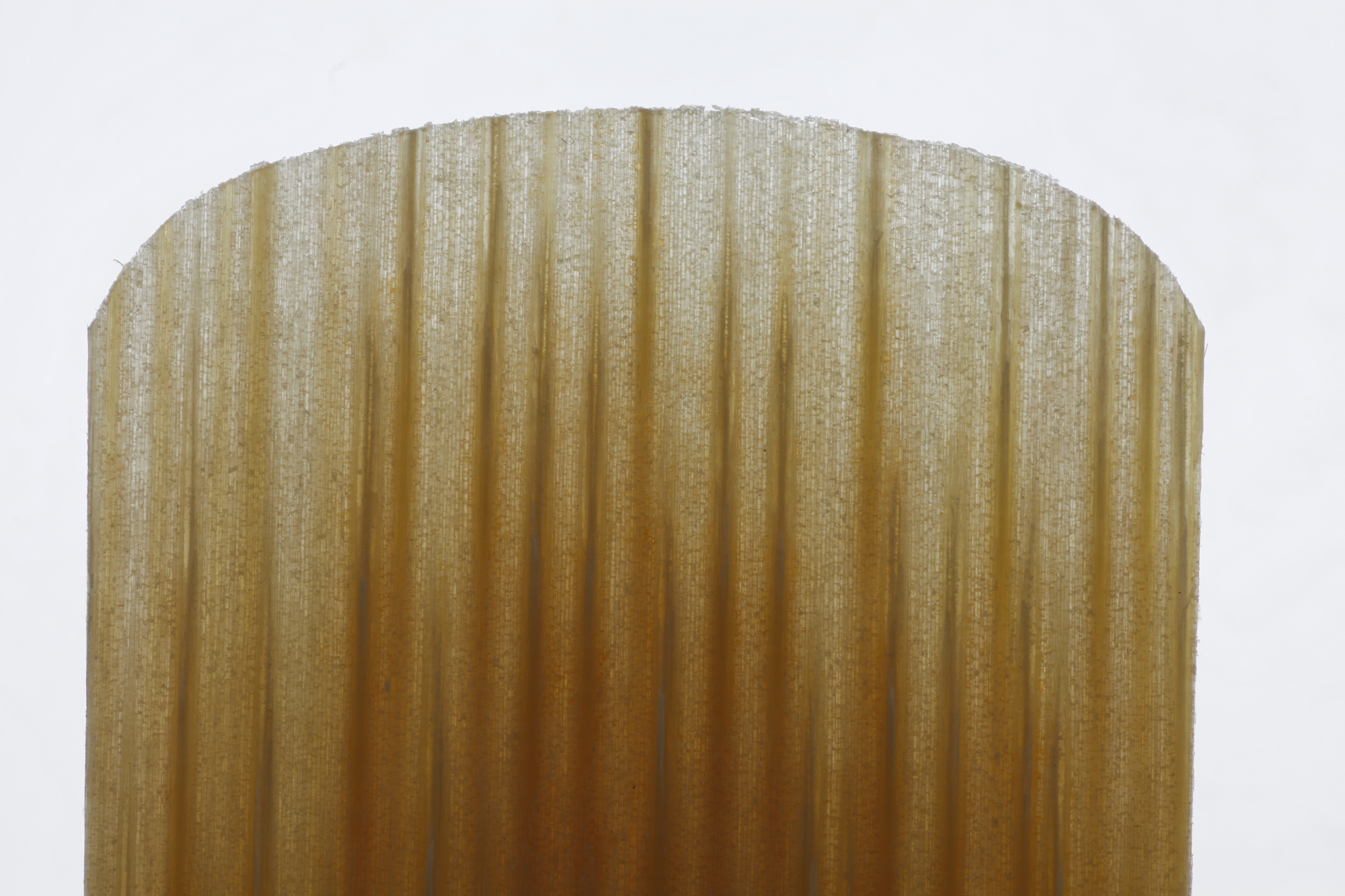
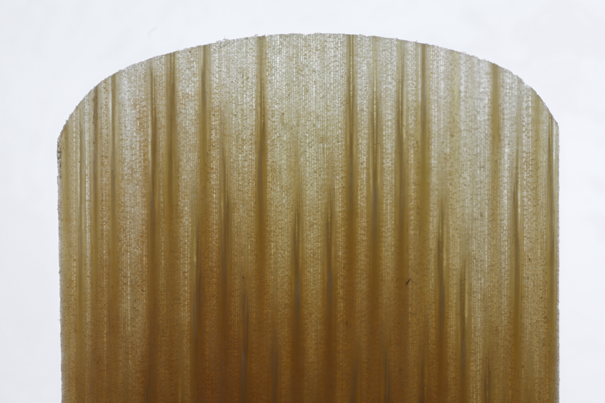
SunnyDaze - your pictures are interesting and thank you for posting them. I was wondering in the photo of the two reeds you posted on the 29th, what to make of the dark strands in the 1 1/2 reed and none in the harder reed?
John Gibson, Founder of JB Linear Music, www.music4woodwinds.com
|
|
Reply To Message
|
|
Author: SunnyDaze
Date: 2019-07-22 21:57
Hi John,
Thanks, I'm glad that you liked them. I wondered about the dark streaks too.
Here's what I think is happening with the streaks:
In the wood there are vascular bundles. The vascular bundles are like drinking straws that conduct water and minerals and suger through the stem of the plant.
The vascular bundles are composed of vertical tubes called xylem and phloem. The xylem conducts water and minerals upwards from the roots to the leaves. The phloem conducts sugar down from the leaves to the roots.
The xylem vessels have tough walls that are reinforced with special stuff called lignin. The phloem is not reinforced with lignin.
Anyway, I would say that in the 1.5 reed the yellow streaks are probably heavily lignified xylem tissue. In the 5 reed there seem to be a lot of vascular bundles but they appear to be less heavily, or at least less unevenly, lignified.
This research paper below says that the quality of a clarinet reed is mostly down to the specific qualities of the vascular bundles and fibre in the plant, so that would seem to agree.
https://www.jstor.org/stable/42764997?Search=yes&resultItemClick=true&searchText=Arundo&searchText=donax&searchUri=%2Faction%2FdoBasicSearch%3FQuery%3DArundo%2Bdonax%2B%26amp%3Bacc%3Doff%26amp%3Bwc%3Don%26amp%3Bfc%3Doff%26amp%3Bgroup%3Dnone&ab_segments=0%2Fdefault-2%2Fcontrol&refreqid=search%3Afc44d995d2e84a6e545faedb473f0e19&seq=1#page_scan_tab_contents
The thing that I wonder now is, would it make sense to take the nice even non-streaky 5 reed and just sand it down a bit until it is thinner and might it then be better then the streaky 1.5? I might need some sandpaper to work that out.
|
|
Reply To Message
|
|
Author: Ken Lagace
Date: 2019-07-22 23:21
>>would it make sense to take the nice even non-streaky 5 reed and
>>just sand it down a bit until it is thinner and might it then be better
>>than the streaky 1.5?
After making hand made reeds for over 50 years, I now get harder reeds and fix them lighter to my taste. They are always better than a virgin reed of the same strength.
|
|
Reply To Message
|
|
Author: clarnibass
Date: 2019-07-23 02:18
Just a suggestion. You see those dark lines and dots, with repeating patterns? Not on the reed, just random areas, mostly in the background. Those are dust and some other dirt on the sensor. It "moves" like this in the stack because the software aligns and matches the subject, so those dust spots move. The subject moves because the camera moves which changes perspective. You can clean them easily with any cloning/healing tool. I imagine your field (research, etc.) might be one where sometimes cloning anything is not allowed? Probably not in this case though.
|
|
Reply To Message
|
|
Author: SunnyDaze
Date: 2019-07-23 04:17
Hi Ken,
That sounds really sensible. The 5 strength reed seems so much more consistent, so that if I could sand it down in the right way then it must be better than the 1.5.
I will start reading around and have a go.
I also would like compare my two favourite reed brands to see what the difference is, as that might give me a clue about where to sand, so I will do that next.
Clarnibass - weirdly the lines are actually bits of fluff on the reeds. I keep a big towel over my setup to keep the dust off and it seems to have added these bits of fluff instead. Ooops!
Sunny
|
|
Reply To Message
|
|
Author: Bob Bernardo
Date: 2019-07-23 06:00
Wish you have emailed me. The information is good, meaning interesting, but it's sadly not going to make a good reed nor figure out the strength differences. For that reason I have to back out. I wanted to write a publication that meant something to the forum.
I had 4 or 5 years of structured studies under microscopes but unable to photograph them. I went as far as using assorted colored dyes and amazing experiments to help figure out cell structures and hardnesses of reeds and the cell walls as well. Also the different countries where the best and worst cane came from. I have to keep my secrets for now. Maybe some other time we can try this.
Best of luck, your information is indeed fun and interesting to the readers. I think this is great!
Glad you are doing this.
Designer of - Vintage 1940 Cicero Mouthpieces and the La Vecchia mouthpieces
Yamaha Artist 2015
|
|
Reply To Message
|
|
Author: SunnyDaze
Date: 2019-07-23 10:46
Hi Bob,
Thanks for commenting again. I completely understand that your work is secret, and that's really fine.
I have written to Vandoren and they said that they are happy for us to do this investigative work publically, so I figure we just start from scratch and have some fun working it out. I've always worked in public science rather than private, so I realise we may be a little way behind, but I think it's nice to be able to put the results in the public domain.
Anyway, we will have a bit of fun looking. :-) Should I still email you? I don't want to trouble you if you have committed to keeping your knowledge secret.
Best wishes,
Sunny
|
|
Reply To Message
|
|
Author: SunnyDaze
Date: 2019-07-24 00:10
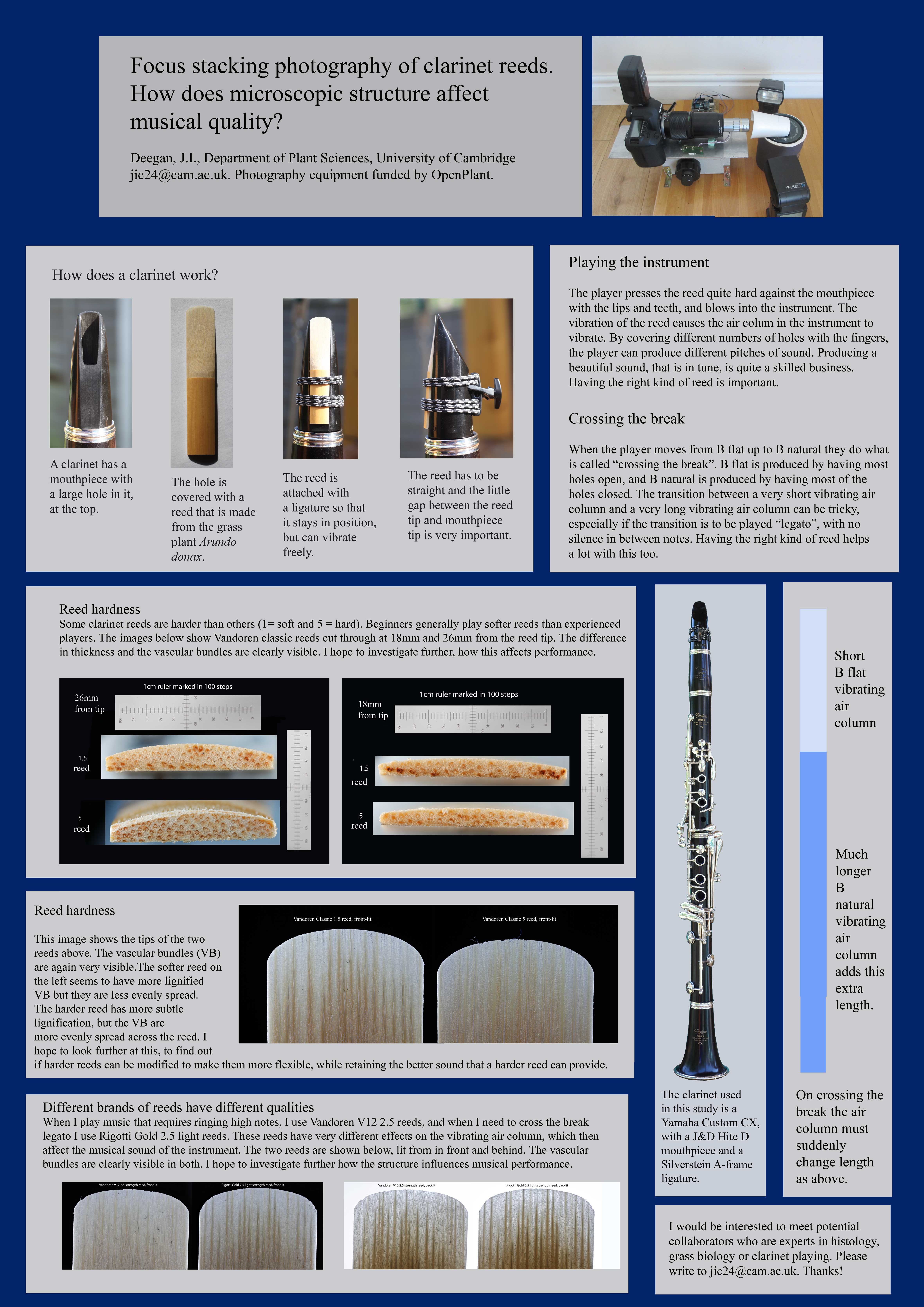
Attachment: poster-very-small.jpg (1772k)
For bonus fun, I am taking a poster about this to the conference of the organisation that paid for my microscope objectives next week. It is a conference full of plant scientists so should be fun. They will be surprised to find me there with a clarinet in my hand, and pleased that I am using my microscope. :-) The poster is attached.
|
|
Reply To Message
|
|
Author: SunnyDaze
Date: 2019-07-24 00:28
Hello :-)
I forgot to say, but would you like me to credit the BBoard community on the author line of the poster? I have done that in the abstract in the conference book, but not yet on the actual poster. Unless anybody objects I will add that credit to the poster too.
|
|
Reply To Message
|
|
Author: Mark Charette
Date: 2019-07-24 03:44
SunnyDaze wrote:
> Yes, good idea. I'll change that. Thanks!
The Clarinet BBoard at Woodwind.Org would be even better
|
|
Reply To Message
|
|
Author: clarnibass
Date: 2019-07-24 08:19
>> weirdly the lines are actually bits of fluff on the reeds. I keep a big towel over my setup to keep the dust off and it seems to have added these bits of fluff instead. Ooops! <<
I didn't mean the stuff on the reeds. I mean some of the dots and smears on the background. They are only visible in the non-cropped photos when the background is bright enough. Their shape is an obvious sign that they are on the sensor and repeating from the alignment when stacking.
|
|
Reply To Message
|
|
Author: SunnyDaze
Date: 2019-07-24 10:02
Oh, gosh. Thanks for mentioning. I'll get it to do a sensor clean and see if that helps. Thanks!
|
|
Reply To Message
|
|
Author: clarnibass
Date: 2019-07-24 12:58
That would help, but it's hard to get it 100% clean. Those Mitutoyo objective have about an f/19 aperture which isn't the worst but shows a lot of dust and dirt. If cloning/healing is allowed for your photos that is the easiest, often I do this before stacking, then paste to all the photos. Since it's on the sensor the location is the same in all the photos.
|
|
Reply To Message
|
|
Author: SunnyDaze
Date: 2019-07-25 22:20
I can't actually see any of the dust spots that you mean. Where is it that you're seeing them?
|
|
Reply To Message
|
|
Author: SunnyDaze
Date: 2019-07-30 18:13
Thanks, I'll have a look at my various images and see if that recurs in other places. I don't think it can be the sensor as it's not on all my images.
|
|
Reply To Message
|
|
Author: SunnyDaze
Date: 2019-07-30 18:19
I just presented my poster at the conference and a guy had a really interesting idea.
He said that he thought porosity of the reed was probably really important and that if a new reed was wetted, then the cells would likely swell up and alter the musical properties of the reed significantly. He said that he has some very serious machinery that could be used to look at the porosity of the reeds before and after wetting, and that he'd be really pleased to have a look if I could send suitable reeds to him.
I thought it was really interesting that he suggested that wetting the reed would have a big effect, as people have mentioned on here that cycles of wetting-drying make a big difference to the musical properties of the reed.
I'm planning to send him some reeds that are new and unused, and some that have had several wet-dry cycles so he can have a look and see what's going on. He's an academic (public funded) scientist so we'd be able to publish the research so everybody could have a look.
I wondered if that would fit with your idea of how reeds work in real life?
Thanks!
|
|
Reply To Message
|
|
Author: SunnyDaze
Date: 2019-08-02 23:58
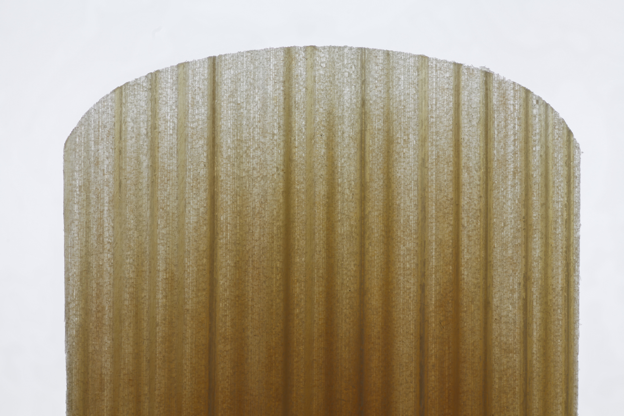
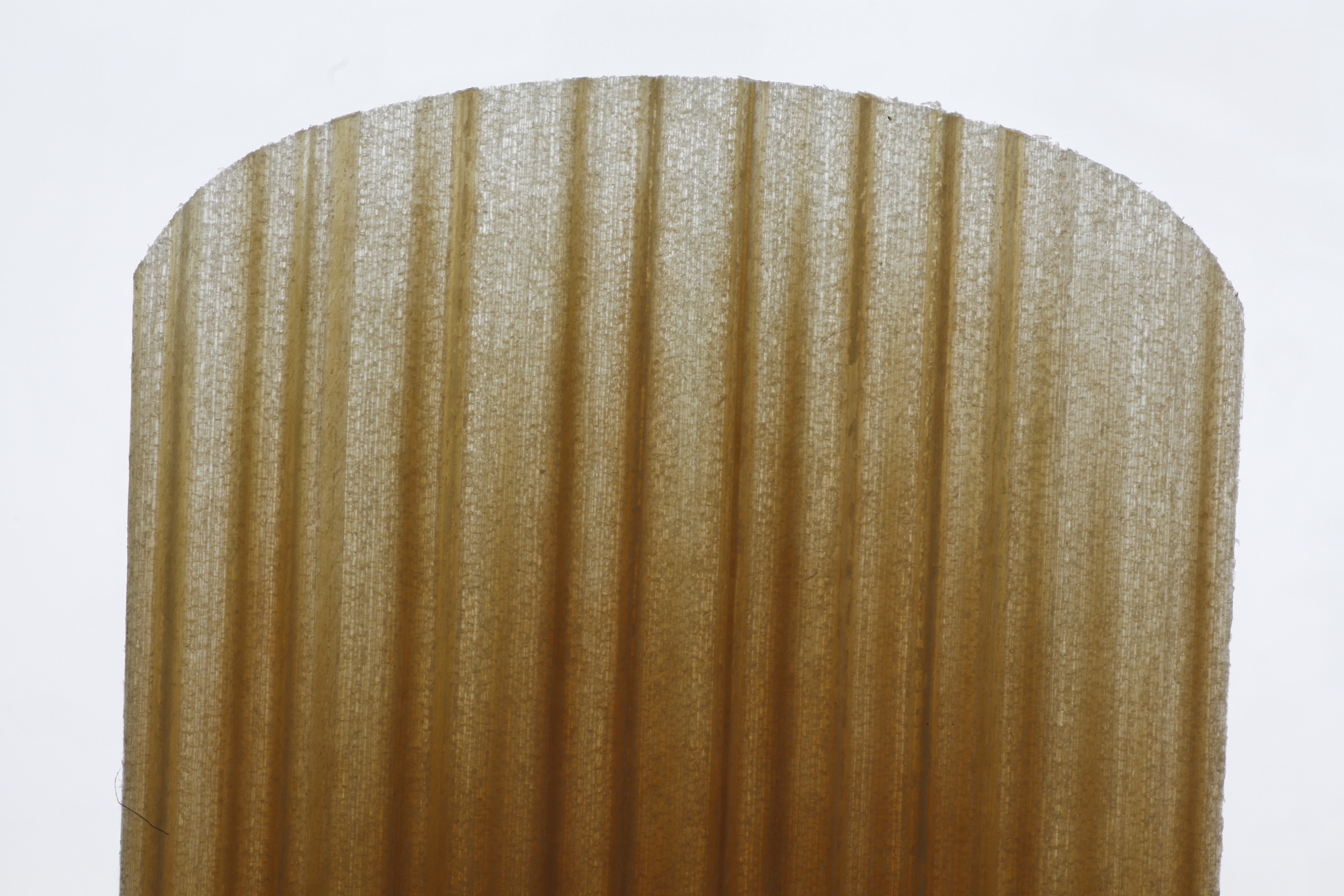
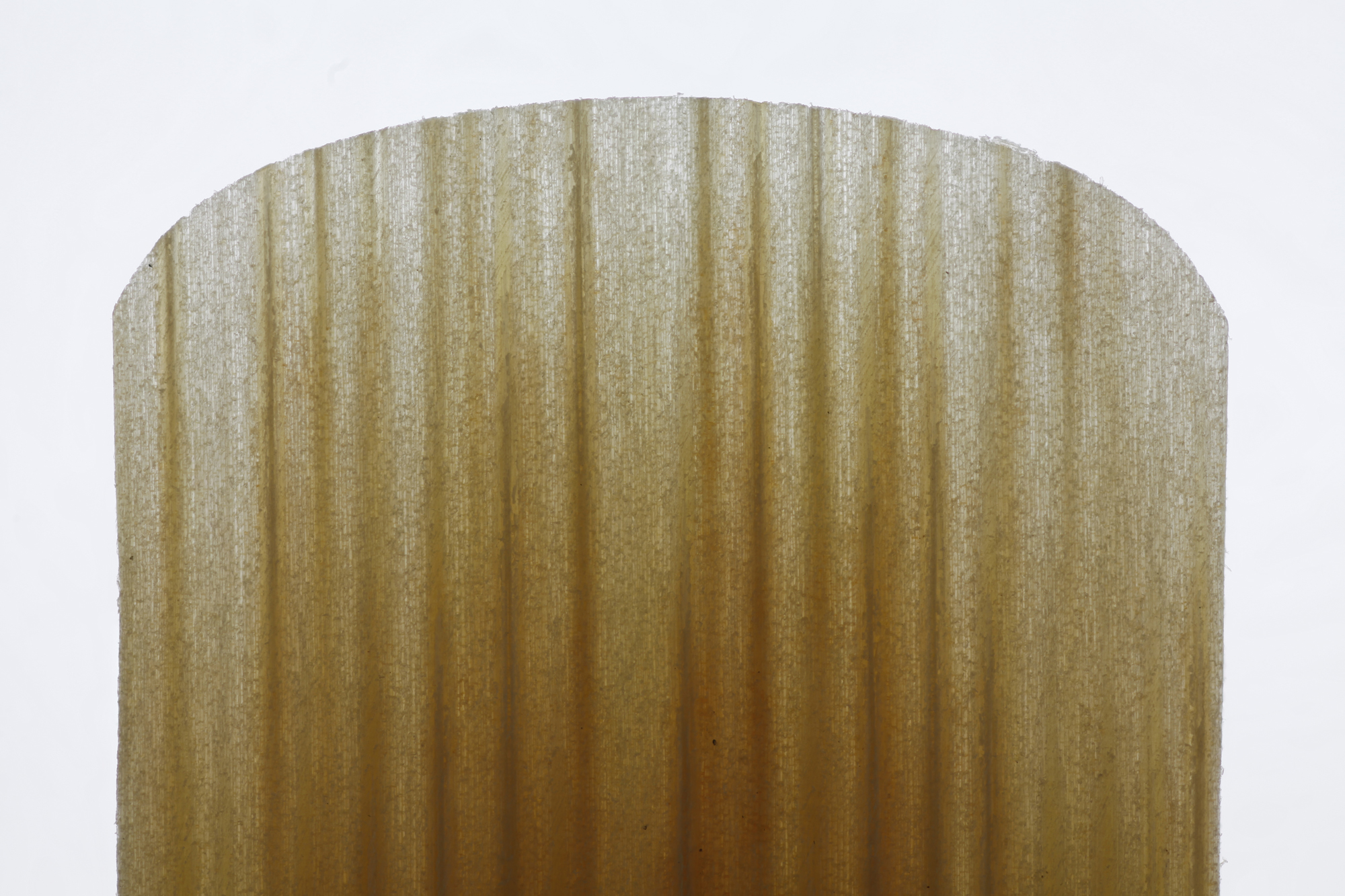
Attachment: 1.jpg (1449k)
Attachment: 2.jpg (1515k)
Attachment: 3.jpg (1462k)
Attachment: 4.jpg (1379k)
Attachment: 5.jpg (1336k)
Hi,
I just took photos of a box of 10 x 2.5 Rigotti Gold light reeds. They're amazingly variable within one box. I wondered if you would like to see? The images are attached here.
Sunny
|
|
Reply To Message
|
|
Author: SunnyDaze
Date: 2019-08-03 00:00
These are the ones where I found the first one that I tried was brilliant for crossing the break so I'm going to try a whole box and see what they're all like, and whether it correlates with the pattern of the lignified areas that are visible.
|
|
Reply To Message
|
|
Author: kdk
Date: 2019-08-04 21:44
I'm not surprised at most of the variability - we're not dealing with a manufactured product, after all. But I am curious how nos. 9 and 10 turn out to play - they seem noticeably darker up into the tip area. I can't tell - maybe you can - if that's a difference in the cut or in the cane itself.
Karl
|
|
Reply To Message
|
|
Author: SunnyDaze
Date: 2019-08-04 23:40
Hi Karl,
Thanks for looking. No. 10 didn't play well at all. It was too hard and had a very hesitant response. Looking at the image, I think it is just very hard because the dark areas are lignifed xylem vessels and it really has lot of them, and heavily lignified. (They are like pieces of wire, reinforcing the reed, so having too many makes it very hard.)
I haven't tried no 9 yet, but I will tomorrow and will report back. I do see what you mean. It looks really well reinforced just like 10.
No 1 is wonderful. I am trying to break in 1 and 5 gently to I can send them off to another microscopist for more analysis. I played 1 again today and it was absolutely brilliant. I tried my tune where I have to cross the break legato, and I'm almost certain that I can do it right on that reed. I haven't made a recording yet to see if the computer agrees, and I don't see my teacher until Thursday. It really felt like the perfect reed though.
I have been looking at very hard Vandoren 5 strength reeds and wondering if I could sand them down really thin to make great reeds. They seem to have such a lot of very fine lines in them. I'm not sure though. I might have a go.
Thanks so much for looking. :-)
|
|
Reply To Message
|
|
Author: kdk
Date: 2019-08-05 02:34
SunnyDaze wrote:
> I have been looking at very hard Vandoren 5 strength reeds and
> wondering if I could sand them down really thin to make great
> reeds.
Most reed adjustment consists of thinning areas to make them vibrate more easily and improve the response. So, empirically, to an extent thinning can compensate for a reed's natural stiffness in a specific location or zone, or for a cut that leaves some area less flexible than the player prefers.
But keep in mind that the strength grade is determined by the cane's stiffness, or so the reed manufacturers tell us, not by the dimensions of the profile, since they are graded after they exit the cutters.
I've never seen the cutting process live - only on videos from Vandoren - but Bob Bernardo certainly has (and probably others), and could probably verify the different strengths are cut to the same nominal specs. The tools I use to measure reeds suggest that all V.12s measure within certain limits regardless of strength, even though any two even of the same strength may not be the same.
As I suggested in my first response, I'm very curious about what measurable differences actually exist (if not in the cut, then in the cane's properties) between a #5 and a #3 or #4 or, for that matter, #1 of the same model reed.
Karl
|
|
Reply To Message
|
|
Author: SunnyDaze
Date: 2019-08-05 11:17
Hi Karl,
Thanks, yes I see what you mean. So finding out what the measurable difference between the actual plant tissue of a #1.5 versus a #3 versus a #5 would be interesting. That's definitely something we could think about.
I have met a researcher who is interested to collaborate, and I could talk to him and see if he has ideas about that. At the moment we have one experiment planned as follows:
I took one box of Rigotti Gold 2.5 light reeds. I photographed them backlit. I chose four good ones (evenly spaced brown lines, and not too brown). I am breaking in two and leaving two unused. I'm planning to post them to my new collaborator and he's going to have a good look at them and see what he can figure out with his equipment. (I have no idea what equipment he has, but I think he's a grass tissue specialist-ish)
Possibly I could change the plan so that I send him just one broken-in reed and one dry one, and then I could also send him a #2.5 and a #5 to compare. That would still be four reeds for him to look at.
Do you think that would be good? I have all those to hand.
Thanks so much for talking to me about this. I really appreciate it.
Sunny (or Jennifer, in real life)
|
|
Reply To Message
|
|
Author: SunnyDaze
Date: 2019-08-05 11:19
Hi again,
I forgot to say - the reason for our other experiment is that my collaborator friend works a lot with grasses and is convinced that porosity of the reed will be a huge factor in it's musical usefulness. He says that he thinks the breaking-in process is probably a lot to do with the water in the mouth gradually changing the porosity of the reed tissue, and he would like to look at that experimentally.
Jen
|
|
Reply To Message
|
|
Author: kdk
Date: 2019-08-05 17:05
SunnyDaze wrote:
> He says that he thinks the breaking-in
> process is probably a lot to do with the water in the mouth
> gradually changing the porosity of the reed tissue, and he
> would like to look at that experimentally.
There's also a lot of guessing among reed players about the role of bacteria and of mineral precipitates in changes to the cane during break-in and over the entire period of use and how this might influence whether to use tap water or saliva for routine wetting.
Karl
|
|
Reply To Message
|
|
Author: SunnyDaze
Date: 2019-08-05 18:22
Hi Karl,
Thanks, that's really interesting to know. I'll ask my friend.
Best wishes,
Jen
|
|
Reply To Message
|
|
Author: kdk
Date: 2019-08-05 18:41
SunnyDaze wrote:
> Possibly I could change the plan so that I send him just one
> broken-in reed and one dry one, and then I could also send him
> a #2.5 and a #5 to compare. That would still be four reeds for
> him to look at.
>
> Do you think that would be good? I have all those to hand.
That sounds good. If his experience with grasses provides any different a point of view, it could be instructive.
Karl
|
|
Reply To Message
|
|
Author: SunnyDaze
Date: 2019-08-05 19:26
Thanks, yes, that was what I was hoping. He just buzzed up to me at that conference I went to, and was really excited about the subject because he has a completely different angle on it. I have no idea what he's going to do, but it will be interesting to find out.
|
|
Reply To Message
|
|
The Clarinet Pages
|
 |
.jpg)
.jpg)
